
It’s just a flesh wound!
Deep carious lesions irritate the pulp and cause its inflammation. In some cases this continues to spread leading to necrosis (death) of the dental pulp and infection of the root canal system. Timely and correct treatment of these carious lesions can avoid the need for more invasive treatments.

A fourteen year old was referred to me as they were experiencing pain from the lower right side. The periapical radiograph shows a deep carious lesion on the mandibular second molar that has breached the mesial pulp horn. It is also clear that there is incomplete root development.
When I saw the patient I did a vitality test for this tooth which confirmed my suspicion that the tooth was still vital. This information decided the treatment, a vital pulp therapy.

All the caries was removed under rubber dam isolation and the pulp was accessed. Inflamed pulp tissue to a depth of a couple of millimetres was removed with sterile instruments and the bleeding was stopped by applying pressure with cotton soaked in sodium hypochlorite.
Mineral trioxide aggregate (MTA) was placed as a direct pulp cap. This has excellent biocompatibility and will allow the pulp to heal. I used white MTA as it does not discolour the tooth. This was given 10 minutes to set then a composite restoration was placed on top.
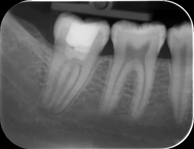
Two years and no symptoms later a review radiograph shows continued root development on the mandibular second molar.
Great result!