
One, good. Two, great. Three, I’m getting excited now!
The root canal morphology of the maxillary first molar can be complex. The teeth tend to have three roots with four canals, the mesio-buccal (MB) root containing two canals. Even though the prevalence of the second MB canal is quite high, 48-97.6% (Martins et al. 2018) finding its elusive orifice is one of the challenges of root canal treatment.
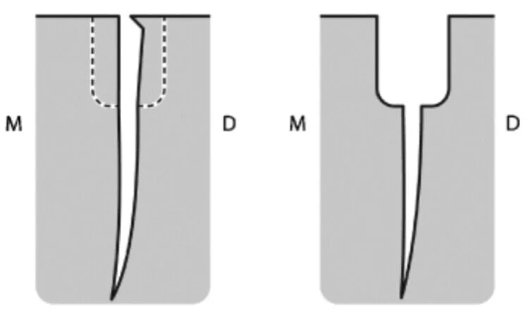
This second MB canal is located in a position mesial to a line drawn between the MB and palatal canal orifices. There is a characteristic lip of dentine covering the orifice which will prevent a handfile from negotiating the full length of the canal. Removing this lip of dentine with ultrasonic tips or goose-neck rose head burs is needed in order to prepare the canal.
Now then, onto an interesting case I was lucky enough to treat.

This patient was in pain due to an infected non-vital maxillary right first molar. He went to see an emergency dentist who got the patient out of pain by accessing the root canal system and dressing the canals with an antibacterial medicament.

After accessing the the pulp chamber I noticed the existing restoration had a well-sealed distal margin and decided to keep this in place as it will help keep the irrigant within the tooth.
After accessing and preparing the main MB canal, I carefully removed dentine to locate the second MB canal. To my surprise I unearthed two extra canal orifices, the second and third MB canals. I mechanically prepared these canals and it became apparent that they both joined the main MB canal towards the apex.
You can see all three MB canal orifices in a line and also the disto-buccal canal orifice above.
The presence of a third MB canal in maxillary first molars can range from 0.5% (Tomaszewska et al. 2018) to 11.3% (Rezaeian et al. 2018).

After preparing and irrigating all the canals, they were dried and obturated with gutta percha using a warm condensation technique.
I need to take an angled radiographic view at the review appointment to really show up the three canals in the MB root.